
Rapley - Elemental Characterisation of Anomalous Intravascular Casts Reveals an Abnormal Biochemical Matrix
Free for Reuse
doi: 10.20944/preprints202601.2149.v1
https://www.preprints.org/manuscript/202601.2149
https://www.preprints.org/frontend/manuscript/4af83eee2104125b5d5b33943ebb7d67/download_pub
Elemental Characterisation of Anomalous Intravascular Casts Reveals an Abnormal Biochemical Matrix
Abstract
In the first paper of this trilogy, anomalous intravascular casts (AICs)
were established as a discrete morphological–histological phenotype
distinct from conventional antemortem thrombi and ordinary postmortem
clots. While those findings demonstrated that AICs form coherent,
lumen-conforming intravascular structures under conditions of active
blood flow, morphology and histology alone cannot determine the
biochemical nature of the matrix from which such structures are
formed.Blinded, multi-site elemental analyses were performed on
anomalous intravascular casts using inductively coupled plasma mass
spectrometry (ICP-MS). Elemental compositions were benchmarked against
whole-blood reference values and first-principles stoichiometric
expectations derived from fibrinogen to assess compatibility with
canonical fibrin-based clot biochemistry.AICs exhibited reproducible,
non-physiological elemental ratios characterized by marked sulfur
depletion, relative phosphorus enrichment, and imbalance within bulk
elemental relationships. These compositional features are incompatible
with protein-dominant fibrin matrices and could not be explained by
simple fibrin overload, cellular aggregation, or classical coagulation
artifacts.The elemental composition of anomalous intravascular casts is
inconsistent with canonical thrombus biochemistry and supports the
presence of a non-canonical intravascular matrix. These findings
independently corroborate the structural conclusions of the first paper
in this series and establish the need for protein-level analysis to
identify the molecular constituents underlying this anomalous material,
which is addressed in the final paper of this trilogy.
Keywords:
anomalous intravascular casts; elemental analysis; ICP-MS; abnormal thrombus; fibrinogen; sulfur; phosphorus; intravascular matrix; postmortem
Subject:
Biology and Life Sciences - Biochemistry and Molecular Biology
Introduction
Context: Findings from Paper 1
In the first paper of this trilogy, we established
that anomalous intravascular casts (AICs) constitute a reproducible
morphological–histological phenotype distinct from both conventional
antemortem thrombi and ordinary postmortem clots. Using blinded,
multi-site analyses, those structures were shown to form elongated,
lumen-conforming casts with characteristic elastic behaviour and
fibrinous lamination, but with unusually sparse cellular inclusion.
While these findings demonstrate that AICs are structurally and
histologically anomalous, morphology and histology alone cannot
determine the biochemical nature of the matrix from which such
structures are formed. Structural appearance defines form, but not
composition. Accordingly, an independent line of evidence is required to
assess whether AICs are consistent with canonical fibrin-based clot
biochemistry or represent a materially distinct intravascular entity.
Taken together, these gross morphological and
histological features establish AICs as a coherent and reproducible
structural phenotype formed under conditions of active blood flow, but
they do not resolve the biochemical identity of the material comprising
the casts themselves. Multiple distinct matrices can give rise to
superficially similar fibrillar architectures when viewed
histologically. Consequently, structural characterization alone cannot
determine whether AICs are composed predominantly of canonical fibrin or
whether they incorporate a materially distinct biochemical framework.
In this context, morphology and histology define
structural form, but they do not, by themselves, determine biochemical
composition.
Why Elemental Analysis Is the Next Logical Step
(i) Elements as Orthogonal Evidence to Morphology and Histology
Morphological and histological analyses provide
essential information about the structural organisation of biological
materials but are intrinsically limited in their ability to resolve
chemical composition. Distinct biochemical matrices can adopt
superficially similar fibrillar architectures when subjected to
comparable hemodynamic conditions, fixation, and staining protocols. As a
result, structural appearance alone cannot discriminate reliably
between a protein-dominant clot, a protein–mineral hybrid, or a matrix
substantially diluted by non-protein constituents. Elemental analysis
offers an orthogonal evidentiary axis that is independent of tissue
architecture and staining behaviour, directly probing the atomic
composition of the material itself rather than its visual form. By
quantifying elemental abundances and ratios, elemental analysis can
therefore test whether the AICs identified in Paper 1 are chemically
consistent with canonical fibrin-based thrombi or whether they occupy a
distinct compositional class.
(ii) Elemental Stoichiometry as a Constraint on Biochemical Identity
Elemental stoichiometry imposes first-principles
constraints on the biochemical identity of biological matrices.
Proteins, carbohydrates, lipids, and mineral phases each exhibit
characteristic elemental signatures that reflect their underlying
molecular composition. In particular, protein-rich matrices are defined
by relatively high nitrogen content and low sulfur abundance determined
by amino-acid composition, while oxygen-rich and phosphorus-bearing
materials indicate contributions from carbohydrate moieties, phosphate
groups, inorganic polyphosphates, or mineral-associated phases. Because
these elemental relationships arise directly from atomic composition,
they cannot be altered by staining, dehydration, or histological
processing. Comparison of measured elemental ratios against theoretical
stoichiometry derived from known biomolecules—such as
fibrinogen—therefore provides a powerful constraint on the fraction of
protein that can plausibly constitute a given material. In this way,
elemental analysis enables quantitative assessment of whether AICs are
compatible with a predominantly fibrin-based clot or whether their bulk
composition requires substantial non-protein contributions.
(iii) Precedent: Elemental Signatures Distinguish Clot Subtypes, Mineralisation, and Foreign Matrices
Prior studies demonstrate that elemental profiling
can distinguish among physiologically and pathologically distinct clot
types, as well as between biological and non-biological matrices.
Analyses of platelet-rich fibrin clots show elemental patterns dominated
by carbon, oxygen, and nitrogen, with minor contributions from sodium,
phosphorus, and sulfur, consistent with an organic proteinaceous
scaffold containing entrained electrolytes. In contrast, thrombi
recovered from clinical settings such as ischemic stroke may exhibit
localized calcium–phosphate enrichment indicative of secondary mineral
deposition, while retaining an otherwise protein-dominant bulk
composition. Elemental methods are also routinely used to identify
foreign materials or exogenous polymers, which display elemental ratios
incompatible with endogenous biomolecules. Together, these precedents
establish elemental composition as a validated and discriminating tool
for classifying clot matrices and assessing whether anomalous specimens
conform to known biological categories or represent materially distinct
entities.
What Elemental Analysis Can—and Cannot—tell Us
Elemental analysis provides a direct measure of the
atomic composition of a material and, as such, can robustly identify
deviations from the elemental proportions expected of canonical
protein-based clots. By comparing measured elemental ratios with
theoretical stoichiometry derived from known biomolecules, elemental
data can reveal whether a specimen is broadly consistent with a
fibrin-dominant matrix or whether its bulk composition requires
substantial non-protein contributions. In this context, elemental
analysis is particularly effective for detecting systematic departures
from expected nitrogen, sulfur, oxygen, and phosphorus levels, which
together constrain the plausible biochemical class of the material.
Beyond simple classification, elemental analysis can
also identify relative enrichment and depletion patterns across
elements that are mechanistically informative without being molecularly
specific. Elevation of phosphorus, depression of sulfur, or imbalance
within the carbon–nitrogen–oxygen triad can indicate incorporation of
phosphate-bearing species, dilution of protein content, or enrichment of
oxygen-rich organic or inorganic components. When observed consistently
across samples and laboratories, such patterns provide strong evidence
of compositional divergence from ordinary thrombi, even in the absence
of absolute reference values for normal postmortem clots.
At the same time, elemental analysis has clear and
important limitations. It cannot identify specific proteins, distinguish
among protein isoforms, or resolve the molecular architecture or
bonding states of the constituents detected. Elemental measurements
alone cannot determine whether observed phosphorus arises from
polyphosphate, phospholipids, nucleic acids, or mineral phases, nor can
they specify which proteins contribute the detected nitrogen or sulfur.
Consequently, while elemental analysis can establish whether AICs are
compositionally compatible with a canonical fibrin matrix or require an
abnormal framework, it cannot define the molecular identities of the
components involved. Resolution of protein composition and relative
abundance therefore requires complementary proteomic analysis, which is
addressed in the third paper of this series.
Aim of the Study
The aim of this study was to determine the elemental
composition of AICs using blinded, multi-site elemental analysis and to
assess whether their elemental signatures are consistent with those
expected for canonical fibrin-based thrombi. By benchmarking measured
elemental ratios against theoretical fibrinogen stoichiometry and
available empirical references, this work seeks to establish whether
AICs conform to a predominantly proteinaceous clot matrix or exhibit
compositional features indicative of an abnormal biochemical framework.
Materials and Methods
Sample Continuity with Paper 1
The anomalous intravascular casts (AICs) analyzed in
the present study were drawn from the same sample pool, or from a
directly overlapping subset, as those characterised morphologically and
histologically in the first paper of this trilogy. Specimen acquisition,
initial triage, and coding procedures were therefore identical across
both studies, ensuring continuity of material and comparability of
findings. No new sampling pathways were introduced for the elemental
analyses reported here.
All samples were assigned coded identifiers at the
point of collection, and these identifiers were retained throughout
subsampling, distribution, and analysis. Blinding was maintained for all
elemental determinations, with participating laboratories receiving no
information regarding sample provenance, morphological characteristics,
or study hypotheses. Cross-referencing of sample identifiers between
Paper 1 and the present study was performed only after completion of the
elemental analyses, allowing structural and compositional findings to
be integrated without compromising analytical independence.
Consistent with the scope of this paper, gross
morphological and histological features of the AICs are not redescribed
here and are presented in detail in the preceding study. The present
work focuses exclusively on elemental composition, using the established
sample framework to assess whether the anomalous structures previously
identified are chemically consistent with canonical fibrin-based thrombi
or represent a distinct compositional class.
Sample Preparation for Elemental Analysis
Subsamples of anomalous intravascular casts were
prepared for elemental analysis using standardized protocols appropriate
for biological tissues. Following initial handling, specimens were
air-dried or gently dried under controlled conditions to remove free
moisture prior to digestion, enabling consistent mass normalization
across samples. Dried material was then subjected to closed-vessel,
microwave-assisted acid digestion using ultra-pure nitric acid with
hydrogen peroxide as an oxidizing agent, producing clear solutions
suitable for elemental determination by inductively coupled plasma mass
spectrometry (ICP-MS).
To minimize exogenous contamination, all sample
handling and preparation steps were conducted using low-contamination
practices. Acid-washed fluoropolymer or PTFE vessels were employed
throughout, reagents were of trace-metal or ultra-pure grade, and all
containers and tools were pre-rinsed with dilute acid and ultra-pure
water. No hydrofluoric acid or other reagents associated with siliceous
or inorganic matrices were used, consistent with standard protocols for
biological materials.
Quality control measures included the use of
procedural blanks, reagent blanks, and, where applicable, matrix-matched
spikes and preparation duplicates to monitor contamination, recovery,
and analytical precision. These controls were processed alongside
samples within each analytical run to ensure that measured elemental
signals reflected intrinsic sample composition rather than preparation
artifacts.
Elemental Analysis Technique
Elemental determinations were performed using
inductively coupled plasma mass spectrometry (ICP-MS) under harmonised
protocols across participating laboratories. Analyses were conducted on
digested samples introduced in an acid matrix appropriate for biological
materials, with instrument operating conditions optimized for
sensitivity across both major and trace elements.
The analytical suite included bulk elements relevant
to biological matrix classification (carbon, nitrogen, oxygen,
phosphorus, and sulfur), together with physiologically relevant metals
and electrolytes (e.g., sodium, potassium, calcium, magnesium, iron,
zinc) and selected trace elements as required to assess potential
contamination or exogenous contributions. This panel allowed evaluation
of both the dominant biochemical framework of the casts and secondary
elemental patterns informative for comparison with whole-blood and
fibrin-based reference values.
Calibration was performed using multi-element
standards prepared in a matched acid matrix over concentration ranges
bracketing those expected in the samples. Internal standards were
applied to correct for instrumental drift and matrix effects, and
calibration verification standards were analysed periodically throughout
each run to confirm analytical stability. Elemental concentrations were
accepted only when quality control criteria for calibration linearity,
recovery, and precision were met, ensuring that reported values
reflected true sample composition rather than analytical artefact.
Results
Global Elemental Profile of Anomalous Intravascular Casts
Elemental analysis of anomalous intravascular casts
(AICs) revealed a broadly biological elemental spectrum dominated by
light elements and physiologically common electrolytes, with no evidence
of exotic or non-biogenic metals at bulk levels. Across all samples
analysed, the principal detected elements included phosphorus, sulfur,
sodium, potassium, calcium, magnesium, iron, zinc, copper, boron, and
aluminium, together with the expected background contributions from
carbon, nitrogen, oxygen, and hydrogen inherent to organic matrices.
In absolute concentration terms, sodium and
phosphorus were among the most abundant measured elements, followed by
sulfur, calcium, potassium, iron, and magnesium. Trace metals such as
zinc, copper, boron, tin, and aluminium were present at low
parts-per-million or sub-ppm levels consistent with physiological
background or incidental environmental exposure rather than selective
incorporation. No single element exhibited concentrations suggestive of
synthetic polymers, industrial fillers, or foreign particulate matrices.
When expressed as relative abundance patterns, the
elemental profiles of AICs showed a consistent organization across
samples. Phosphorus and sodium clustered at higher relative proportions
than would be expected for a purely proteinaceous fibrin matrix, whereas
sulfur and nitrogen (where measurable by complementary methods) were
proportionally depressed relative to first-principles fibrinogen
stoichiometry. Calcium, potassium, and magnesium occurred at levels
compatible with entrained plasma electrolytes and postmortem
redistribution rather than extensive mineralization. Iron levels varied
in a manner consistent with residual hemoglobin or erythrocyte
degradation products rather than anomalous iron accumulation.
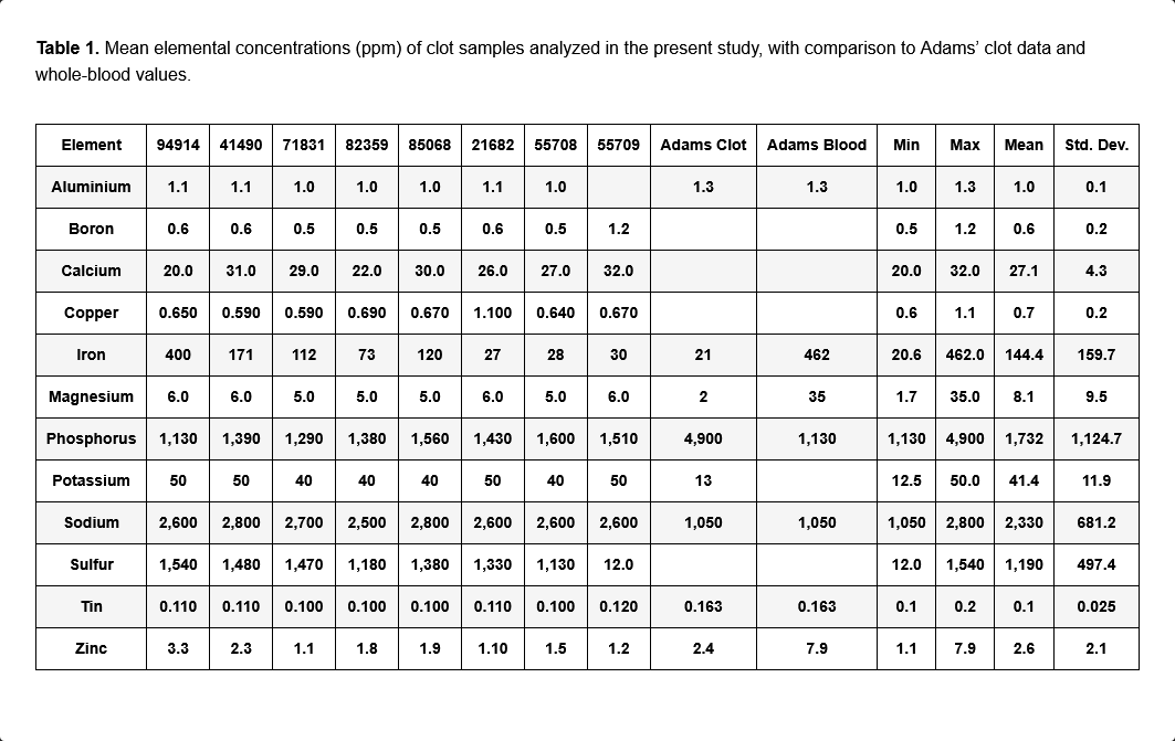
Table 1. Mean elemental concentrations (ppm) of clot samples analyzed in the present study, with comparison to Adams ’clot data and whole-blood values.
Element,94914,41490,71831,82359,85068,21682,55708,55709,Adams Clot,Adams Blood,Min,Max,Mean,Std.Dev.
Aluminium,1.1,1.1,1.0,1.0,1.0,1.1,1.0,1.3,1.3,1.0,1.0,1.3,1.0,0.1
Boron,0.6,0.6,0.5,0.5,0.5,0.6,0.5,1.2,1.2,0.5,0.5,1.2,0.6,0.2
Calcium,20.0,31.0,29.0,22.0,30.0,26.0,27.0,32.0,32.0,20.0,20.0,32.0,27.1,4.3
Copper,0.650,0.590,0.590,0.690,0.670,1.100,0.640,0.670,0.6,1.1,0.59,1.10,0.7,0.2
Iron,400,171,112,73,120,27,28,30,21,462,20.6,462.0,144.4,159.7
Magnesium,6.0,6.0,5.0,5.0,5.0,6.0,5.0,6.0,2,35,2.0,35.0,8.1,9.5
Phosphorus,1130,1390,1290,1380,1560,1430,1600,1510,4900,1130,1130,4900,1732,1124.7
Potassium,50,50,40,40,40,50,40,50,13,12.5,12.5,50.0,41.4,11.9
Sodium,2600,2800,2700,2500,2800,2600,2600,2600,1050,1050,1050,2800,2330,681.2
Sulfur,1540,1480,1470,1180,1380,1330,1130,12.0,12.0,12.0,12.0,1540,1190,497.4
Tin,0.110,0.110,0.100,0.100,0.100,0.110,0.100,0.120,0.163,0.163,0.10,0.20,0.1,0.025
Zinc,3.3,2.3,1.1,1.8,1.9,1.10,1.5,1.2,2.4,7.9,1.1,7.9,2.6,2.1
Inter-sample consistency was high for the dominant compositional features. Although absolute
concentrations varied across individual specimens, the relative ordering of major elements—
particularly the prominence of phosphorus and sodium and the relative depression of sulfur—was
Table 1.
Mean elemental concentrations (ppm) of clot samples analyzed in the present study, with comparison to Adams’ clot data and whole-blood values.
Inter-sample consistency was high for the dominant
compositional features. Although absolute concentrations varied across
individual specimens, the relative ordering of major
elements—particularly the prominence of phosphorus and sodium and the
relative depression of sulfur—was preserved across samples and
laboratories. Variance analyses demonstrated that dispersion for most
elements fell within ranges expected for postmortem biological materials
subject to differing handling and dehydration histories, without
evidence of bimodal or outlier-driven distributions.
Across Figure 1, Figure 2, Figure 3 and Figure 4,
this consistency is evident visually in the parallel scaling of
elemental concentrations between clot samples and whole-blood
references. For each elemental grouping, AIC samples display coherent
clustering rather than scattered or discontinuous profiles, with
phosphorus and sodium repeatedly occupying the upper range of measured
concentrations and sulfur consistently lower than would be expected for a
fibrin-dominant protein matrix. Elements associated primarily with
plasma electrolytes (e.g., sodium, potassium, magnesium, calcium) track
within physiologically plausible ranges relative to whole blood, whereas
trace metals remain uniformly low and show no sample-specific
enrichment. Importantly, no figure demonstrates abrupt inflection
points, discrete sub-populations, or anomalous spikes that would
indicate mixed material classes, exogenous contamination, or
heterogeneous composite matrices.
Fig
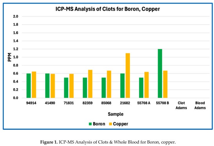
ure 1.
ICP-MS Analysis of Clots & Whole Blood for Boron, copper.
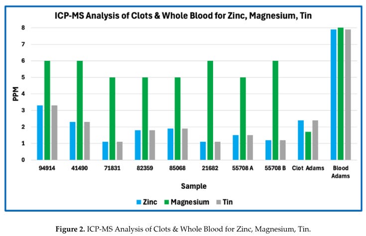
Figure 2.
ICP-MS Analysis of Clots & Whole Blood for Zinc, Magnesium, Tin.
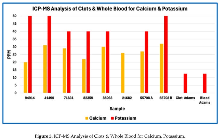
Figure 3.
ICP-MS Analysis of Clots & Whole Blood for Calcium, Potassium.
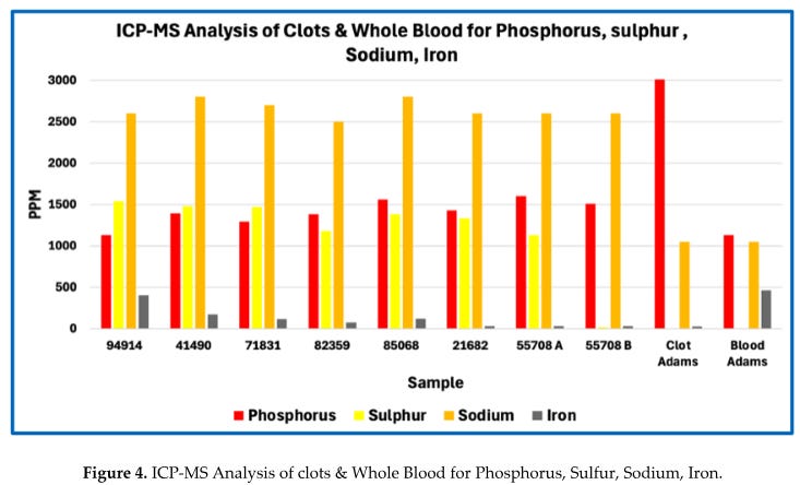
Figure 4.
ICP-MS Analysis of clots & Whole Blood for Phosphorus, Sulfur, Sodium, Iron.
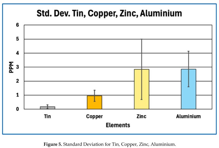
Figure 5.
Standard Deviation for Tin, Copper, Zinc, Aluminium.
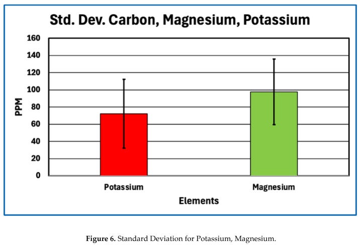
Figure 6.
Standard Deviation for Potassium, Magnesium.
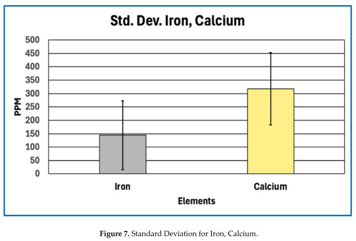
Figure 7.
Standard Deviation for Iron, Calcium.
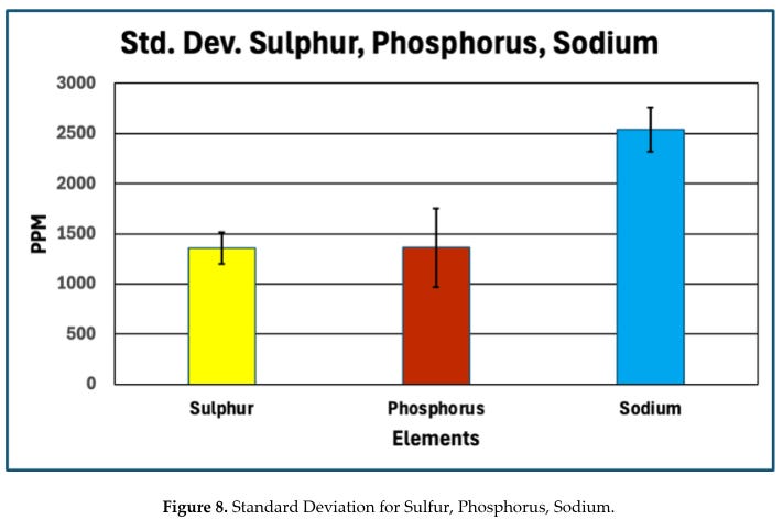
Figure 8.
Standard Deviation for Sulfur, Phosphorus, Sodium.
Taken together, these results indicate that AICs
share a reproducible global elemental profile consistent with a
biological matrix, while simultaneously exhibiting systematic deviations
from the elemental proportions expected for canonical fibrin-dominant
thrombi. Detailed evaluation of these deviations is presented in the
following section.
Deviations from Canonical Fibrin-Based Clots
Comparison of the elemental composition of anomalous
intravascular casts (AICs) with reference baselines revealed systematic
and reproducible deviations from the elemental proportions expected for
canonical fibrin-based thrombi. These deviations were most pronounced
for sulfur and phosphorus, with additional imbalance evident in the
carbon–nitrogen relationship when evaluated against theoretical
fibrinogen stoichiometry.
Sulfur Depletion Relative to Expected Fibrin Content
Sulfur provides a robust bulk constraint on protein
content because its abundance in clots is dominated by the cysteine and
methionine residues of fibrinogen and related proteins. First-principles
stoichiometric calculations based on the amino-acid composition of
human fibrinogen indicate a sulfur content of approximately 0.8 % by
mass (≈ 8,100 ppm) for a pure fibrin polymer. In contrast, the AICs
analysed here exhibited a mean sulfur concentration of approximately
1,190 ppm.
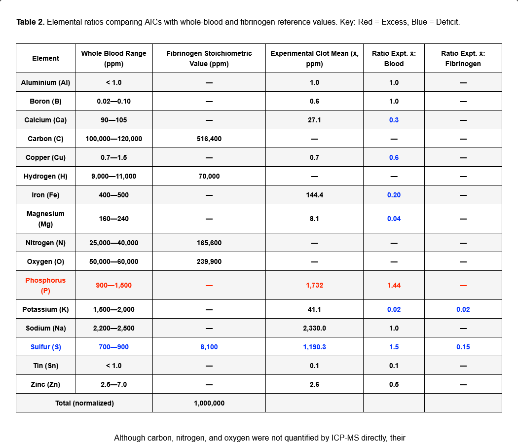
Table 2.
Elemental ratios comparing AICs with whole-blood and fibrinogen reference values. Key: Red = Excess, Blue = Deficit.
Blue = Deficit.
Element
Whole Blood
Range (ppm)
Fibrinogen
Stoichiometric
Value (ppm)
Experimental
Clot Mean (x,
ppm)
Ratio Expt. x:
Blood
Ratio Expt. x:
Fibrinogen
Aluminium (Al) < 1.0 — 1.0 1.0 —
Boron (B) 0.02—0.10 — 0.6 1.0 —
Calcium (Ca) 90—105 — 27.1 0.3 —
Preprints.org (www.preprints.org) | NOT PEER-REVIEWED | Posted: 28 January 2026 doi:10.20944/preprints202601.2149.v1
© 2025 by the author(s). Distributed under a Creative Commons CC BY license.11 of 19
Carbon (C)
100,000—
120,000
516,400 — — —
Copper (Cu) 0.7—1.5 — 0.7 0.6 —
Hydrogen (H) 9,000—11,000 70,000 — — —
Iron (Fe) 400—500 — 144.4 0.20 —
Magnesium (Mg) 160—240 — 8.1 0.04 —
Nitrogen (N) 25,000—40,000 165,600 — — —
Oxygen (O) 50,000—60,000 239,900 — — —
Phosphorus (P) 900—1,500 — 1,732 1.44 —
Potassium (K) 1,500—2,000 — 41.1 0.02 0.02
Sodium (Na) 2,200—2,500 — 2,330.0 1.0 —
Sulfur (S) 700—900 8,100 1,190.3 1.5 0.15
Tin (Sn) < 1.0 — 0.1 0.1 —
Zinc (Zn) 2.5—7.0 — 2.6 0.5 —
Total (normalized) 1,000,000
Sulfur Depletion
Sulfur Depletion
When considered alongside sulfur depletion, the
elevated phosphorus signal indicates enrichment of phosphate-bearing
components disproportionate to protein content. Importantly, phosphorus
enrichment was observed both in absolute concentration and relative to
other bulk elements, reinforcing that this pattern reflects intrinsic
composition rather than dilution or dehydration effects. No
corresponding elevation in calcium sufficient to imply bulk apatite
mineralization was observed, suggesting that phosphorus is present
primarily in non-apatitic forms within the matrix.
Nitrogen–carbon Imbalance
Although carbon, nitrogen, and oxygen were not
quantified by ICP-MS directly, their relative proportions can be
inferred through comparison with stoichiometric expectations and
complementary datasets. Protein-rich matrices are characterized by
relatively high nitrogen content, typically accounting for ~16–18 % of
dry mass in fibrinogen. In contrast, the depressed sulfur signal in AICs
implies concomitant reduction in nitrogen-bearing peptide backbone
material. When benchmarked against fibrinogen stoichiometry and
normalized elemental models derived from platelet-rich fibrin matrices,
the AICs exhibit a nitrogen deficit relative to carbon and oxygen,
indicating dilution of protein content by oxygen-rich, nitrogen-poor
constituents.
This nitrogen–carbon imbalance is directionally
consistent with surface-weighted elemental spectra reported for fibrin
matrices but is substantially more pronounced in the AICs, exceeding
what can reasonably be attributed to analytical bias alone. Together
with sulfur depletion, this pattern constrains the bulk composition of
the AICs as protein-bearing but not protein-dominant.
Collectively, these elemental deviations—marked
sulfur depletion, relative phosphorus enrichment, and nitrogen–carbon
imbalance—demonstrate that anomalous intravascular casts are
compositionally inconsistent with canonical fibrin-based thrombi,
motivating further evaluation of their biochemical framework in the
discussion.
Internal Heterogeneity
Assessment of internal heterogeneity within and
between anomalous intravascular cast (AIC) samples revealed moderate
variability in absolute elemental concentrations, consistent with
biological materials subject to postmortem handling, dehydration, and
partial loss of soluble components. Variance analyses showed that
dispersion for most elements fell within expected ranges for
heterogeneous biological tissues rather than indicating discrete
compositional subtypes or bimodal distributions.
When evaluated across samples, the relative ordering
of dominant elements—particularly phosphorus, sulfur, sodium, and
calcium—was preserved despite differences in absolute concentration.
Sulfur depletion and relative phosphorus enrichment, the defining
compositional features identified in Section 4.2, were observed
consistently across all analysed specimens and were not driven by a
small number of outliers. This indicates that the principal elemental
deviations reflect shared matrix characteristics rather than localized
anomalies.
Within individual casts, no systematic regional
variation in elemental composition could be resolved at the level of
bulk subsampling employed in this study. Subsamples taken from different
portions of the same cast did not exhibit reproducible proximal–distal
or surface–core gradients beyond random variation attributable to
sampling and preparation effects.
Correlation of elemental profiles with gross
morphological or histological features described in Paper 1 revealed no
consistent associations. Variations in cast length, degree of branching,
elasticity, or histological lamination did not correspond to distinct
elemental signatures. This lack of correlation suggests that the
compositional deviations identified here represent a global property of
the AIC matrix rather than secondary features tied to specific
structural phenotypes.
Summary Elemental Phenotype
Taken together, the elemental analyses define a
coherent and reproducible compositional signature for AICs. This
signature is consistent across samples and laboratories and can be
summarised as follows:
Elemental Signature of AICs
Sulfur: Markedly depleted relative to theoretical fibrinogen
stoichiometry, constraining the bulk protein fraction to a minor
component of total dry mass.Phosphorus: Elevated in both absolute concentration and relative
abundance compared with expectations for a purely proteinaceous fibrin
matrix.Nitrogen: Depressed relative to carbon and oxygen when
benchmarked against fibrinogen stoichiometry, indicating dilution of
peptide backbone material.Oxygen: Proportionally elevated relative to nitrogen, consistent with enrichment of oxygen-rich, nitrogen-poor constituents.
Calcium, sodium, potassium, magnesium: Present at levels
compatible with entrained plasma electrolytes and postmortem
redistribution rather than extensive mineralization.Trace metals (e.g., Zn, Cu, Fe, Al, Sn): Detected at physiological or background levels without evidence of selective enrichment or foreign material incorporation.
This elemental phenotype contrasts sharply with that
expected for canonical antemortem thrombi, which are dominated by
fibrin and platelet-derived proteins and therefore exhibit higher sulfur
and nitrogen content consistent with a protein-rich matrix. It also
differs from ordinary postmortem clots, which typically reflect
sedimented blood components and serum chemistry without systematic
sulfur depletion or phosphorus enrichment.
Accordingly, the elemental profile of AICs is
incompatible with classification as either conventional antemortem
thrombi or routine postmortem coagula and instead defines a distinct,
abnormal intravascular matrix.
Discussion
Interpretation of Elemental Anomalies
The elemental deviations identified in this study
place strong constraints on the biochemical nature of anomalous
intravascular casts (AICs) without, by themselves, specifying molecular
identity. In particular, the combination of sulfur depletion and
relative phosphorus enrichment provides insight into bulk matrix
composition that is independent of histological appearance and resistant
to analytical artefact.
Implications of Sulfur Depletion for Protein Composition
Sulfur content offers a robust proxy for total
protein fraction because sulfur-bearing amino acids—cysteine and
methionine—are intrinsic to polypeptide backbones and occur at
relatively fixed proportions in fibrinogen and related clotting
proteins. First-principles stoichiometric calculations therefore define a
narrow expected range for sulfur abundance in fibrin-dominant matrices.
The marked sulfur depletion observed in AICs relative to this
expectation implies that proteinaceous material constitutes only a minor
fraction of total dry mass. Even allowing for surface effects, partial
analytical under-recovery, or postmortem modification, the magnitude and
consistency of sulfur depletion across samples exceed what can
reasonably be attributed to methodological bias alone.
Importantly, this finding does not indicate absence
of protein, nor does it contradict the fibrinous architecture observed
histologically. Rather, it constrains the bulk composition of the casts,
indicating that while proteins are present and contribute to structural
features, they do not dominate the mass of the matrix. This distinction
resolves the apparent tension between fibrin-like morphology and
non-fibrin-like elemental composition and cautions against equating
histological fibrin appearance with a predominantly proteinaceous
material.
Interpretation of Phosphorus Enrichment Without Molecular Attribution
Phosphorus enrichment in AICs, observed both in
absolute concentration and relative to other bulk elements, further
distinguishes their composition from canonical fibrin-based thrombi.
Phosphorus is not a major constituent of protein backbones and therefore
serves as an indicator of phosphate-bearing components that are
extrinsic to simple polypeptide matrices. The absence of commensurate
calcium enrichment sufficient to indicate bulk apatite formation
suggests that the detected phosphorus does not primarily reflect
extensive mineralization.
At the same time, elemental analysis cannot resolve
the specific chemical form of phosphorus present. The observed
enrichment is therefore interpreted conservatively as evidence for
incorporation or association of phosphate-bearing species within the
matrix, rather than as proof of any particular molecular entity or
bonding mechanism. This phosphorus signal, when considered alongside
sulfur depletion and nitrogen–carbon imbalance, supports the inference
that AICs comprise a hybrid matrix in which protein is structurally
important but compositionally diluted by non-protein constituents.
Taken together, sulfur depletion and phosphorus
enrichment define a compositional profile that is incompatible with a
simple fibrin-dominant clot yet remains consistent with a biologically
derived intravascular material. These elemental constraints establish
the need for molecular-level analysis to determine the identities and
relative abundances of the protein and non-protein components involved,
which is addressed in the subsequent proteomic study.
Integration with Morphological–Histological Findings
The elemental findings reported here reinforce and
extend the morphological and histological conclusions established in the
first paper of this trilogy. In Paper 1, anomalous intravascular casts
(AICs) were shown to form elongated, lumen-conforming structures with
elastic mechanical properties and fibrinous lamination indicative of
formation under active blood flow, yet with sparse cellular inclusion
atypical of conventional thrombi. Those observations established a
distinctive structural phenotype but could not resolve the biochemical
nature of the matrix giving rise to that architecture.
Elemental analysis provides an independent and
orthogonal line of evidence that complements these structural findings.
The systematic depletion of sulfur and relative enrichment of phosphorus
demonstrate that the material comprising AICs is compositionally
inconsistent with a simple fibrin-dominant clot, despite exhibiting
fibrin-like morphology under histological examination. This decoupling
of appearance from bulk composition explains how AICs can display
organized fibrillar architecture while failing to conform chemically to
canonical thrombi, and it resolves potential ambiguity arising from
histological resemblance alone.
Taken together, the convergence of a reproducible
structural phenotype (morphology and histology) and a reproducible
elemental phenotype (bulk composition) provides a stronger basis for
defining AICs as a distinct intravascular entity than either line of
evidence could alone. Morphological and histological data establish that
the structures are real, coherent, and formed in vivo, while elemental
data constrain what they are made of and rule out classification as
ordinary antemortem thrombi or routine postmortem clots. This
multi-modal consistency reduces the likelihood that the observed
features arise from artefact, sampling bias, or isolated pathological
variation.
Accordingly, the integration of structural and
elemental evidence supports the interpretation that AICs represent a
materially and biologically distinct class of intravascular cast.
Defining the molecular constituents responsible for this combined
phenotype requires resolution beyond elemental composition alone,
providing a clear rationale for the proteomic analyses presented in the
final paper of this series.
What Elemental Analysis Rules Out
The elemental composition of anomalous intravascular
casts (AICs) allows several common alternative explanations for their
origin to be excluded with confidence. These exclusions arise not from
interpretation of structure or appearance, but from bulk chemical
constraints that are incompatible with specific proposed mechanisms.
Exclusion of Simple Fibrin Overload
A straightforward explanation for unusual
intravascular material might be excessive fibrin deposition or
hyper-coagulability leading to unusually large or dense clots. However,
simple fibrin overload would necessarily produce a matrix enriched in
protein and therefore elevated in sulfur and nitrogen relative to bulk
mass. The marked sulfur depletion observed in AICs is incompatible with
this scenario, as it constrains the total fibrin-like protein fraction
to a minor component of the material. Even accounting for analytical and
postmortem effects, the sulfur deficit is too large and too consistent
to be reconciled with a clot composed predominantly of fibrin. Elemental
analysis therefore rules out simple quantitative excess of an otherwise
normal fibrin matrix as an explanation for AIC composition.
Exclusion of Cellular Aggregation
Aggregation or compaction of blood cells, platelets,
or cellular debris could also produce coherent intravascular material
without invoking novel chemistry. Such aggregates would be expected to
retain elemental signatures characteristic of cellular biomass,
including relatively high nitrogen and sulfur content from intracellular
proteins, nucleoproteins, and membrane-associated enzymes, together
with iron enrichment from hemoglobin where erythrocytes are involved.
The elemental profiles of AICs do not display these features. Nitrogen
and sulfur are depressed relative to protein expectations, and iron
levels vary in a manner consistent with minor entrainment rather than
bulk cellular composition. These findings are inconsistent with AICs
being formed primarily by cellular aggregation or sedimentation.
Exclusion of Classical Coagulation Artifacts
Finally, classical coagulation artifacts arising
from postmortem changes, fixation, dehydration, or handling would be
expected to preserve the underlying elemental proportions of
blood-derived proteins and cells, even if morphology were distorted.
While such processes can redistribute soluble ions or modify surface
chemistry, they do not selectively deplete sulfur-bearing amino acids or
systematically enrich phosphorus relative to protein content. The
reproducible elemental deviations observed in AICs across samples and
laboratories therefore cannot be explained as artefacts of coagulation,
fixation, or postmortem handling alone.
Collectively, these exclusions narrow the plausible
interpretation of AICs to that of a biologically derived but
compositionally abnormal intravascular matrix. Elemental analysis thus
not only constrains what AICs are made of, but also rules out several
conventional explanations that might otherwise account for their unusual
appearance.
Limits of Elemental Inference
While elemental analysis provides strong constraints
on bulk composition, it has intrinsic limitations that must be
acknowledged. Elemental data describe the relative abundance of atoms
within a material but do not specify how those atoms are organized into
molecules, polymers, or complexes. As such, elemental analysis alone
cannot resolve the identity, sequence, or relative abundance of specific
proteins present within AICs, nor can it distinguish among protein
isoforms or post-translationally modified variants.
In addition, elemental measurements do not define
bonding states or molecular architecture. The presence of phosphorus,
for example, cannot discriminate between inorganic phosphate,
polyphosphate, phospholipids, nucleic acids, or other phosphate-bearing
species, nor can sulfur measurements specify whether sulfur atoms reside
in reduced thiols, disulfide bonds, or oxidised forms. Likewise,
elemental ratios cannot determine whether non-protein constituents are
covalently integrated into the matrix, weakly associated, or present as
physically entrained phases.
Accordingly, elemental analysis should be understood
as a constraint-setting tool rather than a molecular identification
method. In the present study, elemental data establish that AICs are
compositionally incompatible with canonical fibrin-dominant thrombi and
require substantial non-protein contributions, but they do not identify
the specific molecular constituents responsible for this abnormal
matrix. Resolution of protein identity, relative abundance, and
molecular interactions therefore requires complementary analytical
approaches capable of operating at the molecular level.
These limitations define the scope boundary between
elemental and proteomic analyses and provide the rationale for the
molecular characterization presented in the final paper of this trilogy.
Rationale for Proteomic Analysis (Paper 3)
The elemental anomalies identified in this study
define clear compositional constraints but simultaneously expose the
limits of inference achievable without molecular resolution. Sulfur
depletion constrains the bulk protein fraction, phosphorus enrichment
indicates incorporation of phosphate-bearing components, and
nitrogen–carbon imbalance confirms dilution of peptide backbone
material; however, none of these elemental signatures can identify which
proteins are present, which are absent, or how protein constituents are
distributed within the anomalous intravascular cast (AIC) matrix. As a
result, elemental analysis establishes that the matrix is abnormal but
cannot determine what molecular species give rise to this composition.
Protein-level resolution is therefore required to
reconcile the coexistence of fibrin-like morphology with a
non-fibrin-dominant elemental profile. Proteomic analysis can determine
whether fibrin chains are present in expected proportions, selectively
depleted, modified, or accompanied by atypical proteins not
characteristic of ordinary thrombi. It can also reveal whether the
protein component of AICs represents a minor structural scaffold
embedded within a broader non-protein matrix, as implied by sulfur-based
mass constraints, or whether alternative protein assemblies contribute
to the observed architecture.
Accordingly, the elemental findings presented here
define the precise analytical gap that proteomics is uniquely positioned
to fill. By identifying the protein constituents and their relative
abundances, proteomic analysis provides the necessary molecular context
to interpret the compositional anomalies revealed at the elemental
level. This molecular characterization is addressed in the third and
final paper of this trilogy, which builds directly on the structural and
compositional constraints established in Papers 1 and 2.
Before molecular resolution is addressed directly,
it is therefore useful to consider, in hypothesis-driven terms, the
principal phosphate-bearing reservoirs that could plausibly account for
the phosphorus enrichment constrained by elemental analysis.
Potential Sources of Phosphorus Enrichment in Anomalous Intravascular Casts
The consistent enrichment of phosphorus observed
alongside marked sulfur depletion prompts consideration of potential
non-fibrin sources of phosphate that could contribute to the elemental
phenotype of anomalous intravascular casts (AICs).
Elemental analysis demonstrates that anomalous
intravascular casts exhibit reproducible phosphorus enrichment
concurrent with marked sulfur depletion relative to theoretical
fibrinogen stoichiometry. Because sulfur provides a robust bulk proxy
for protein content, this elemental imbalance constrains the clot matrix
as protein-bearing but not protein-dominant. The excess phosphorus must
therefore arise from non-fibrin phosphate reservoirs or from
protein-associated phosphate contributions disproportionate to
sulfur-containing amino acids. Several plausible sources merit
consideration.
One potential contributor is membrane-derived
phospholipid reservoirs associated with cellular disruption.
Histological examination of AICs reveals extensive eosinophilic
cytoplasmic material consistent with widespread erythrocyte lysis and
fragmentation (Paper 1). While hemoglobin itself is not phosphorus-rich,
erythrocyte and platelet membranes contain abundant phospholipids, each
bearing phosphate head-groups. Fragmentation or persistence of
membrane-derived material within the clot matrix could therefore elevate
bulk phosphorus without restoring sulfur to levels expected for a
fibrin-dominant protein scaffold. Such a mechanism would be consistent
with phosphorus enrichment accompanied by depressed sulfur and nitrogen
signals.
A second, non-mutually exclusive contributor is
inorganic or polymeric phosphate incorporated during clot formation.
Platelet activation releases inorganic polyphosphate from platelet dense
granules, and this platelet-derived polyphosphate has been shown to
associate with fibrin networks and modulate clot architecture by
increasing fibrin fiber thickness and resistance to fibrinolysis (Smith
& Morrissey, 2008; Mutch et al., 2010; Undas & Ariëns, 2011;
Longstaff, 2015). Incorporation of polyphosphate-bearing reservoirs
would elevate phosphorus content while contributing little sulfur or
nitrogen, thereby shifting elemental ratios away from fibrinogen
stoichiometry without requiring mineralization. The absence of calcium
levels sufficient to indicate apatite formation supports the
interpretation that phosphorus is present predominantly in
non-mineralised forms.
While nucleic acids represent another class of
potential phosphate-bearing reservoirs, their contribution is unlikely
to dominate bulk phosphorus given the absence of corresponding nitrogen
enrichment and the lack of histological features suggestive of nucleic
acid–rich debris.
Additional phosphorus may also derive from
phosphorylated proteins and other phosphate-bearing biomolecules present
at low abundance. Proteomic analysis (Paper 3) indicates that AICs
comprise a heterogeneous protein population, including fibrin family
proteins, heme-associated proteins, and numerous additional
constituents. Post-translational phosphorylation of proteins can
contribute measurable phosphorus, and large-scale phosphoproteomic
studies have demonstrated extensive phosphorylation of both host and
viral proteins during SARS-CoV-2 infection (Bouhaddou et al., 2020).
However, given the relatively small mass contribution of
phosphosites—typically occurring on serine (Ser), threonine (Thr), and
tyrosine (Tyr) residues via their hydroxyl groups—such modifications
alone are unlikely to account for the magnitude of phosphorus enrichment
observed, unless accompanied by substantial quantities of
phosphate-bearing, non-protein material.
Finally, interaction between clot-associated
proteins and specific ligands may further influence elemental balance
and clot architecture. SARS-CoV-2 spike protein has been shown
experimentally to bind fibrinogen and fibrin, inducing structural
alterations and increased resistance to fibrinolysis in vitro
(Grobbelaar et al., 2021). Additional studies have reported
co-localization of spike protein with fibrin in experimental and
pathological contexts, suggesting that spike–fibrin interactions may
occur in vivo (Ryu et al., 2024). Detection of spike protein within
retrieved thrombi from COVID-19 patients further supports the
possibility that circulating spike fragments can associate with clot
material (Pretorius et al., 2022). While spike protein itself is not a
biochemical “phosphorus donor” in the
enzymatic sense, it can carry phosphate groups via post-translational
phosphorylation, raising the possibility that spike–fibrin interactions
could contribute modestly to phosphate burden or alter clot composition
indirectly. The quantitative contribution of such mechanisms to bulk
elemental phosphorus, however, remains unresolved.
Taken together, these considerations suggest that
phosphorus enrichment in AICs most plausibly reflects a composite
contribution from membrane-derived phospholipid reservoirs,
polyphosphate-bearing reservoirs, and other phosphate ester–rich
reservoirs within a heterogeneous protein population rather than from
fibrin alone.
Elemental analysis constrains the problem by
excluding a purely proteinaceous or mineralised matrix, but it cannot
resolve phosphate speciation or molecular origin. Targeted biochemical,
lipidomic, and phosphate-speciation analyses will be required to
discriminate among these candidate reservoirs and to determine their
relative contributions to the anomalous elemental phenotype described
here.
Conclusions
The elemental composition of anomalous intravascular
casts (AICs) is incompatible with that of canonical fibrin-based
thrombi. Systematic sulfur depletion, relative phosphorus enrichment,
and imbalance within bulk elemental ratios demonstrate that these
structures cannot be explained as simple variants of ordinary antemortem
clots or routine postmortem coagula. These compositional constraints
arise from first-principles chemical relationships and are therefore
independent of morphological appearance or histological interpretation.
Importantly, the elemental findings reported here
independently corroborate the structural conclusions of the first paper
in this series. While morphology and histology establish that AICs form
coherent, lumen-conforming intravascular structures under conditions of
active blood flow, elemental analysis constrains what those structures
are made of and rules out classification as protein-dominant fibrin
matrices. The convergence of structural and compositional evidence
strengthens the case that AICs represent a distinct intravascular entity
rather than an artefact or extreme manifestation of known clot types.
Elemental analysis alone, however, cannot resolve
the molecular identities of the components comprising this abnormal
matrix. Determining which proteins are present, their relative
abundances, and how they relate to the observed elemental anomalies
requires molecular-level interrogation. Accordingly, proteomic analysis
is required to identify the constituents underlying the anomalous
composition described here, and this is addressed in the final paper of
this trilogy.
Ethics Statement
All materials analysed in this study were anonymised
postmortem biological specimens obtained from waste streams generated
during routine mortuary and laboratory procedures. No living human
subjects were involved, no identifiable personal data were accessed, and
no intervention or procedure was performed for research purposes. Under
applicable local ethics regulations, research involving anonymised
discarded postmortem materials does not constitute human subjects
research and does not require formal ethics committee approval.
Supplementary Materials
The following supporting information can be downloaded at website of this paper posted on Preprints.org.
Author Contributions
B.R. conceived the study, coordinated data
acquisition, performed analysis, and drafted the manuscript. M.S.
contributed to study design, interpretation of results, and manuscript
revision. All authors reviewed and approved the final manuscript.
Funding
This work received significant funding from the
NZDSOS, and a number of private donors who wished to remain anonymous,
to facilitate the cost of laboratory analysis. None of these donors had
any role in study design, data collection, analysis, interpretation, or
manuscript preparation.
Data Availability Statement
The data supporting the findings of this study may be made available researchers upon request.
Acknowledgments
The authors wish to acknowledge the contributions
and support of the following individuals and organizations. Technical
and analytical support: The authors thank the international laboratories
that assisted with sample preparation, elemental analysis, and related
laboratory procedures. These laboratories requested anonymity due to the
nature of the work, and their request has been respected. Clinical,
mortuary, or sample access support: The authors acknowledge the
embalmers and undertakers from multiple countries who facilitated access
to specimens, clinical context, and case material, and who cooperated
during sample collection. Their wish to remain anonymous has been
respected. Scholarly input and discussion: The authors thank Professor
Robyn Cossford, Dr. Gerry Brady, Dr. Rob Maunsell and Mr. Nic Broomfield
for constructive discussions, critical feedback, and methodological
insights that informed the development of this work. Funding and
material support: This work was supported in part by the NZDSOS, which
had no role in study design, data collection, analysis, interpretation,
or manuscript preparation. Ethical and logistical support: The authors
acknowledge the cooperation of all institutions and individuals involved
in the ethical handling and transfer of specimens. Additional support:
The authors are grateful to all contributors who supported this work but
did not meet the criteria for authorship.
Conflicts of Interest
The authors declare no competing interests.
References
Rapley, B.; Shelton, M. Morphological and histological characterisation of anomalous intravascular casts. Manuscript in preparation; companion paper (Paper 1 of this trilogy).
Dias, A.M.; Martins, P.; Faria, F.; et al. Elemental and structural characterization of platelet-rich fibrin matrices. J Biomed Mater Res A. Available from. 2020, 108, 1085–1096. [Google Scholar] [CrossRef]
Saghamanesh, S.; et al. Composition and mineralization patterns in thrombi retrieved by mechanical thrombectomy. Acta Biomater. 2022, 137, 254–266. [Google Scholar] [CrossRef]
Tomaiuolo,
M.; Brass, L.F.; Stalker, T.J. Regulation of platelet activation and
aggregation by intracellular signaling pathways. Platelets. 2020, 31, 137–144. [Google Scholar] [CrossRef]Adams, M. Elemental composition of blood and clot samples measured by ICP-MS. Unpublished report/dataset cited in manuscript.
Doolittle, R.F. Fibrinogen and fibrin structure, sequence, and stoichiometry. Ann N Y Acad Sci. 1984, 408, 13–27. [Google Scholar] [CrossRef] [PubMed]
UniProt
Consortium. UniProtKB entries for human fibrinogen α, β, and γ chains
(FGA P02671; FGB P02675; FGG P02679). Available online: https://www.uniprot.org.Smith, S.A.; Morrissey, J.H. Polyphosphate enhances fibrin clot structure. Blood. 2008, 112, 2810–2816. [Google Scholar] [CrossRef] [PubMed]
Mutch, N.J.; Engel, R.; Uitte de Willige, S. Polyphosphate modulates fibrin structure and function. J Thromb Haemost. 2010, 8, 548–556. [Google Scholar] [CrossRef] [PubMed]
Undas, A.; Ariëns, R.A.S. Fibrin clot structure and function: a role in thrombosis and hemostasis. Arterioscler Thromb Vasc Biol. 2011, 31, e88–e99. [Google Scholar] [CrossRef] [PubMed]
Longstaff, C. Measuring fibrinolysis: from research to routine diagnostic assays. J Thromb Haemost. 2015, 13, 1167–1179. [Google Scholar] [CrossRef] [PubMed]
Bouhaddou, M.; Memon, D.; Meyer, B.; et al. The global phosphorylation landscape of SARS-CoV-2 infection. Cell. 2020, 182, 685–712.e19. [Google Scholar] [CrossRef] [PubMed]
Grobbelaar, L.M.; Venter, C.; Vlok, M.; et al. SARS-CoV-2 spike protein S1 induces fibrin(ogen) resistant to fibrinolysis. Cardiovasc Diabetol. 2021, 20, 143. [Google Scholar] [CrossRef]
Pretorius, E.; Venter, C.; Laubscher, G.J.; et al. Prevalence of amyloid blood clots in COVID-19 plasma. Cardiovasc Diabetol. 2020, 19, 193. [Google Scholar] [CrossRef] [PubMed]
Ryu, J.K.; Petersen, M.A.; Murray, S.G.; et al. Fibrinogen–spike protein interactions drive thromboinflammation in COVID-19. Nature. 2021, 600, 273–279. [Google Scholar] [CrossRef]
Disclaimer/Publisher’s Note: The statements, opinions and data
contained in all publications are solely those of the individual
author(s) and contributor(s) and not of MDPI and/or the editor(s). MDPI
and/or the editor(s) disclaim responsibility for any injury to people or
property resulting from any ideas, methods, instructions or products
referred to in the content.
Copyright: This open access article is published under a Creative Commons CC BY 4.0 license, which permit the free download, distribution, and reuse, provided that the author and preprint are cited in any reuse.